Now Accepting Medicaid At All Locations!
Now Accepting Medicaid At All Locations!

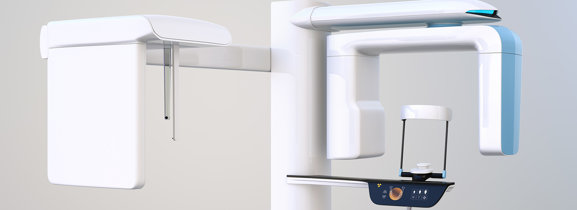
At the office of New Day Dentistry, we rely on modern diagnostic tools to deliver careful, predictable dental care. One of the most important advances in oral imaging is cone-beam computed tomography (CBCT), which captures three-dimensional views of the teeth, jaws, and surrounding structures. These high-resolution images help clinicians see details that traditional two-dimensional X-rays can miss, so treatment decisions are better informed and more precise.
CBCT is a valuable part of our diagnostic toolkit because it provides targeted, distortion-free visualization without requiring invasive procedures. The scan is quick and noninvasive, and the data it produces can be used throughout the course of treatment — from initial evaluation to surgical planning and follow-up. Below, we explain how CBCT works, when it’s most useful, and what patients can expect when this technology is part of their care.
CBCT uses a cone-shaped X-ray beam and a rotating detector to capture a series of images from multiple angles. These projections are then reconstructed by specialized software into a three-dimensional dataset that represents the patient’s anatomy in fine detail. Unlike panoramic or periapical X-rays, which compress complex structures into flat images, CBCT preserves depth and spatial relationships — an essential feature when precise measurements and spatial awareness are required.
The ability to examine anatomy slice-by-slice, rotate views, and zoom into areas of interest enables clinicians to identify problems that can be difficult to detect on standard films. For example, the exact position of an impacted tooth, the proximity of a nerve to a planned implant site, or the extent of a cyst or lesion becomes much clearer. This level of visualization translates to more confident diagnoses and better-informed treatment planning.
In everyday practice, CBCT is not a replacement for all traditional X-rays. Rather, it complements them. Two-dimensional radiographs remain useful for routine screenings and monitoring, while CBCT is reserved for cases where additional three-dimensional detail will change the approach to care. The decision to use CBCT is based on clinical need and the anticipated benefit for diagnosis or treatment.
Dental implant planning is one of the most common and impactful uses of CBCT. The three-dimensional dataset reveals bone volume and density, identifies vital structures like the inferior alveolar nerve and maxillary sinus, and helps determine optimal implant size and position. With this information, clinicians can plan implants with a higher degree of predictability and reduce the risk of complications during surgery.
Endodontic care and the assessment of complex root anatomy also benefit from CBCT’s clarity. Cases with persistent symptoms, unusual canal configurations, suspected root fractures, or periapical pathology often require a level of detail that only three-dimensional imaging provides. CBCT helps pinpoint the exact source of a problem so that treatment — whether non-surgical root canal therapy or surgical intervention — can be targeted and effective.
Additional applications include evaluation of impacted teeth, assessment of jaw pathology or trauma, planning for corrective jaw surgery, TMJ analysis, and airway assessment for sleep-related breathing concerns. In each scenario, CBCT supplies information that directly supports clinical decision-making and improves the chances of a successful outcome.
Concerns about radiation are understandable, and modern CBCT systems are designed with dose efficiency in mind. Compared with older computed tomography (CT) technology, dental CBCT typically uses a lower radiation dose and can be focused on a small field of view to limit exposure to the area of interest. Clinicians follow established guidelines to ensure scans are justified and optimized for each patient’s needs.
The scanning process itself is brief and straightforward. Patients sit or stand while the machine rotates around the head for a matter of seconds to capture the data. There is no contrast material or invasive step required, and most people find the experience comfortable. If a patient is pregnant, highly anxious, or has special medical considerations, the dental team will discuss alternatives and make decisions that prioritize safety.
After the scan, the images are reviewed digitally and can be shared among specialists when multidisciplinary input is needed. Because the dataset is rich and manipulable, clinicians can take precise measurements, create cross-sectional views, and export files for guided surgical planning — all of which contribute to safer, more predictable care.
One of the practical benefits of CBCT is its integration with digital treatment workflows. For implant cases, CBCT data can be combined with intraoral scans to design surgical guides that position implants exactly as planned. This reduces intraoperative guesswork and can shorten procedure time while improving accuracy. For complex reconstructive or orthodontic cases, three-dimensional planning helps visualize outcomes and set realistic expectations.
In restorative and interdisciplinary care, CBCT supports communication between the dentist, lab technicians, and specialists. When restoration margins, root positions, or bone contours affect the final result, having three-dimensional reference material makes coordination more efficient and precise. The end result is a treatment pathway informed by objective data rather than inference.
Beyond planning, CBCT serves as a baseline for monitoring healing and evaluating postoperative results. Follow-up imaging — when clinically appropriate — can demonstrate osseointegration of implants, resolution of pathology, or changes in anatomical relationships over time. This continuity of data supports long-term decision-making and continuity of care.
Accurate interpretation of CBCT scans requires specific training and experience. Our clinical team pairs advanced imaging technology with professional expertise to ensure findings are assessed in the context of the patient’s history and clinical exam. When specialized review is needed, scans can be evaluated in consultation with oral and maxillofacial radiologists or other relevant specialists to provide a comprehensive perspective.
Quality assurance is another important element: routine calibration, appropriate exposure settings, and adherence to imaging protocols help maintain consistent, diagnostic-quality images. Staff training emphasizes patient positioning, reduced motion artifact, and proper selection of field of view to minimize unnecessary exposure while capturing the information essential for care.
Because imaging is a tool that supports clinical judgment rather than replacing it, images are always interpreted in partnership with the patient’s symptoms, exam findings, and treatment goals. This balanced approach helps ensure that CBCT contributes meaningfully to safe, effective dental care.
In summary, cone-beam computed tomography is a powerful diagnostic resource that enhances clarity, improves treatment planning, and supports high-quality clinical outcomes. When used judiciously and interpreted by trained professionals, CBCT offers significant advantages for a wide range of dental procedures while maintaining a focus on patient safety and comfort. If you’d like to learn more about how CBCT might be used in your care, please contact us for additional information.
CBCT is a specialized dental imaging method that captures three-dimensional views of the teeth, jaws and surrounding structures using a cone-shaped X-ray beam and a rotating detector. The machine acquires multiple projections in a single rotation and software reconstructs those images into a detailed 3D dataset. This approach preserves depth and spatial relationships that are lost in conventional two-dimensional radiographs.
The resulting dataset allows clinicians to examine anatomy slice by slice, rotate views and measure structures precisely, which improves diagnostic confidence. Because the scan focuses on a targeted field of view, it provides detailed anatomical information without invasive procedures.
Conventional X-rays such as periapical or panoramic images produce flat, two-dimensional representations of complex anatomy, which can obscure depth and spatial relationships. CBCT preserves three-dimensional detail, enabling clinicians to see the exact position and orientation of teeth, bone contours and nearby vital structures. This difference matters most in cases where precise measurements and spatial awareness change clinical decisions.
That said, CBCT complements rather than replaces standard radiographs; two-dimensional films remain valuable for routine screening and monitoring while CBCT is reserved for cases where additional 3D detail will directly influence care. The choice to use CBCT is based on clinical need and the expected diagnostic benefit.
A dentist may recommend CBCT when three-dimensional information is required to plan or refine treatment. Common indications include dental implant planning, evaluation of complex root anatomy, assessment of impacted teeth, investigation of suspected pathology or fractures, and analysis of the temporomandibular joint or airway.
The recommendation is guided by whether the additional information will change the diagnosis or treatment approach, and clinicians weigh the expected benefit against the need to limit radiation exposure. If CBCT is indicated, the team will explain how the scan fits into the overall care plan.
Modern dental CBCT systems are designed with dose efficiency in mind and typically use lower radiation than conventional medical CT scans; they can also be confined to a small field of view to limit exposure to the area of interest. Clinicians follow established guidelines to ensure that scans are justified and optimized for each patient. Equipment selection, appropriate exposure settings and careful patient positioning help minimize dose while maintaining diagnostic image quality.
Certain situations, such as pregnancy or specific medical conditions, require additional consideration and discussion with the dental team. When scans are performed, providers take steps to ensure patient comfort and safety and will offer alternatives if CBCT is not appropriate.
The CBCT scan is brief and noninvasive; patients typically sit or stand while the scanner rotates around the head for a matter of seconds to capture the dataset. There is no need for contrast agents or invasive preparation, and most people find the process comfortable. Staff will help position you and provide simple instructions to hold still during the acquisition to reduce motion artifact.
After the scan, the images are reconstructed digitally and reviewed by the clinician, and the data can be exported or shared with specialists if multidisciplinary input is needed. The team will discuss the findings with you and explain how the images inform the recommended treatment.
CBCT provides critical information for implant planning by revealing bone volume, bone quality and the precise location of vital anatomical structures such as the inferior alveolar nerve and maxillary sinus. With this three-dimensional view, clinicians can determine optimal implant size, angulation and position while avoiding adjacent anatomy. This level of detail reduces intraoperative uncertainty and supports safer surgery.
CBCT data can also be integrated with intraoral scans and digital planning software to design surgical guides that translate the virtual plan into accurate clinical placement. This digital workflow improves predictability and helps set realistic expectations for the procedure and outcome.
Yes. CBCT is particularly useful in endodontics for identifying complex root canal anatomy, locating additional canals, detecting root fractures and evaluating periapical pathology that may not be visible on two-dimensional films. In cases with persistent symptoms or unclear findings, three-dimensional imaging helps pinpoint the source of pain or infection. This clarity supports targeted treatment whether the approach is nonsurgical root canal therapy or surgical intervention.
By providing precise localization and anatomy visualization, CBCT aids clinicians in planning access, anticipating procedural challenges and monitoring treatment outcomes. When necessary, images can be reviewed in consultation with endodontic specialists to ensure comprehensive care.
Interpretation of CBCT scans requires specific training and experience beyond general radiography, and dentists who use this technology receive education in image analysis, anatomy and potential pitfalls such as artifacts. Depending on the complexity of the case, providers may consult oral and maxillofacial radiologists or other specialists for a formal interpretation. This collaborative approach ensures that findings are placed in the appropriate clinical context.
Quality assurance practices, including routine calibration, protocol adherence and staff training in patient positioning, help produce consistent, diagnostic images. Images are always interpreted alongside the patient’s history and clinical examination to support balanced clinical judgment.
CBCT can provide three-dimensional views of the upper airway and surrounding skeletal structures, which helps identify anatomical contributors to airway restriction such as narrow regions, mandibular position or tonsillar enlargement. These images are useful for visualizing airway volume and spatial relationships that may relate to sleep-disordered breathing. However, CBCT captures static anatomy and does not replace functional tests like polysomnography.
When airway assessment is part of the evaluation, CBCT findings are combined with a comprehensive sleep and medical history and may be discussed with sleep medicine specialists or otolaryngologists. This multidisciplinary perspective helps determine whether imaging findings correlate with symptoms and whether further diagnostic testing or referral is needed. At New Day Dentistry, we coordinate care with appropriate specialists when airway concerns arise.
CBCT data can be merged with intraoral scans and CAD/CAM software to create comprehensive digital treatment plans for restorative, orthodontic and surgical procedures. This integration enables precise design of restorations, prosthetics and surgical guides, and it facilitates communication between the dentist, laboratory technicians and specialists. The result is a coordinated workflow grounded in objective 3D data rather than estimation.
Beyond planning, CBCT serves as a baseline for postoperative assessment and long-term monitoring, allowing clinicians to evaluate healing, osseointegration or changes in anatomy over time. Exportable datasets support efficient collaboration and continuity of care across the dental team.